Wednesday, November 16, 2016

Wellness Day Project and Reflection

Our Wellness Day presentation can be seen here: Wellness Day Presentation

Reflection:

First of all, I chose the topic of tai chi because I felt that it met all of the requirements of this project: it was something that I was familiar with but also interested to try learning about and executing myself, as well as a topic that I felt was obscure enough that my peers could learn a lot about. My father has practiced this type of martial arts since I was young, and watching him do it has always been fascinating to me, so I decided to take a shot at trying it out and seeing for myself how it really was. I had touched on the topic of tai chi during school only once before, in a middle school PE unit, and having forgotten really what was taught during that time, I felt that Wellness Day was a perfect time to revisit that time and bond with my dad even more. Some interesting things that I learned while preparing was that, as I mentioned during our presentation, tai chi is indeed way harder than it looks. It took at least probably 5 hours of practice for Michelle and I to memorize the small portion of the 24-movement tai chi sequence that we used in our activity, and I was able to gain newfound appreciation and admiration for my dad and other people who practice this kind of martial arts. Not only was tai chi difficult to memorize, but the physical taxation of doing it strangely fascinating to me. I was also surprised at the ability that only a small amount of tai chi in my life could calm me; even after our short stint during the Wellness Day activity, I felt infinitely more relaxed and focused than I would be during any other Anatomy & Physiology class after lunch, when I usually feel drowsy. On the topic of general health and wellness that tai chi provides, there are numerous studies proving the long–term mental and physical benefits of practicing this art. Despite the misconceptions of being effortless and straightforward, tai chi has been known to improve the general balance, flexibility and strength of an individual with its almost painfully slow and deliberate movements, and in a way, not using external equipment produces an entirely different challenge in itself. Instead of relying on machines and tools to stretch and strengthen muscles and bones, one has to achieve seemingly impossible positions while looking smooth and graceful all the while. In terms of alleviating certain illnesses, tai chi reduces the risk of, in some cases, arthritis, osteoporosis, high blood pressure, and muscle atrophy in older adults, whereas practicing from a young age not only helps people stay ahead of the game in terms of health, but also assists in improving their attention span. Usually, people who practice tai chi find a connection between the mind and body, and use tai chi in a way similar to meditation or mindfulness, which we learned in class, to focus their minds and concentrate on their breathing and movements. The breathing aspect of tai chi is consequently hugely beneficial to the inducement of the parasympathetic nervous system, as the controlled breathing causes the sympathetic nervous system and feelings of stress to subside. I feel that I would give myself a 9.5/10 because I believe that the effort that I put into our project was quite a lot, and the balance of work between me and Michelle was mostly even. In the future, I think I would be interested to learn more about how long it takes to fully master tai chi, and what other “practical” defenses can be found in many of the movements of tai chi, since I found it really interesting to learn from my dad where the “Parting the Wild Horse’s Mane” move really came from.
Tuesday, November 8, 2016

Unit 3 Reflection

Themes and Essential Understandings


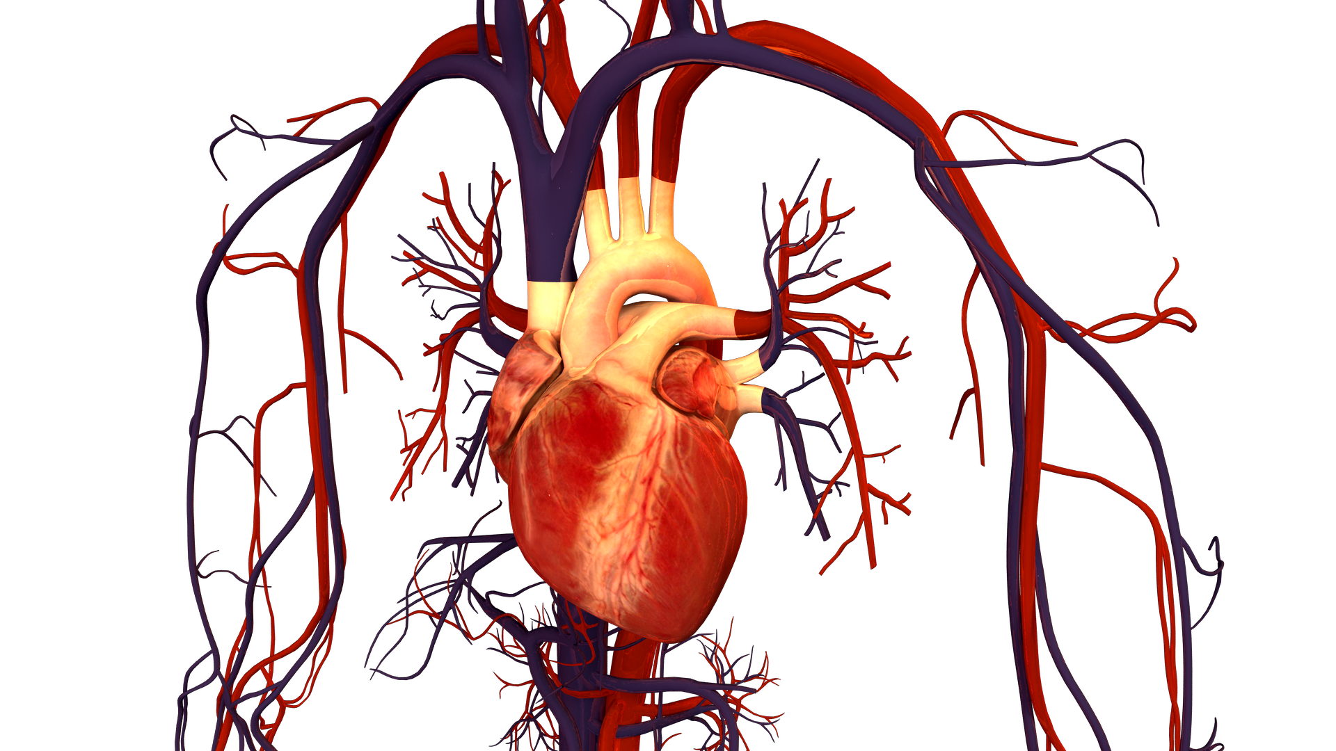
This unit is basically about the importance of cardiovascular health, and aspects of its parts that help it carry out its various functions. The cardiovascular system first starts off with the heart, which pumps blood throughout the body to transport oxygen. Entering through the superior and inferior vena cava to pass through the right atrium and tricuspid valve to the right ventricle, deoxygenated blood makes its way finally out of the heart through the pulmonary artery to the lungs to pick up more  oxygen. On its way back to the parts of the body that need it, the reinvigorated blood travels from the pulmonary veins to the left atria, and passes through the bicuspid valve to go into the left ventricle, where it then exits through the aortic valve and aorta to go to other places in the body.
Brandenburg, Bryan. Human Heart and Circulatory System. Digital image. Wikipedia. N.p., 18 Jan. 2013. Web. 8 Nov. 2016.
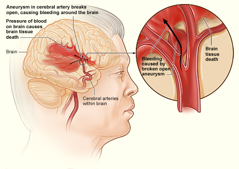
A related system is the respiratory system, where air enters through the nose and mouth to travel down to the lungs, where the bronchus branches out into various bronchioles that later transition into being alveoli that exchanges carbon dioxide with oxygen. Cardiovascular health is the ability for the heart and blood vessels to work efficiently in transporting needed materials, so that the body's processes of consuming oxygen and defending against foreign invaders are not inhibited. Heart disease and stroke are often caused by unhealthy diets with too much sodium and unrestricted carbohydrates and alcohol, as well as a lack of exercise and obesity.
National Heart Lung and Blood Institute. Stroke Hemorrhagic. Digital image. Wikipedia. N.p., 12 Nov. 2013. Web. 8 Nov. 2016.
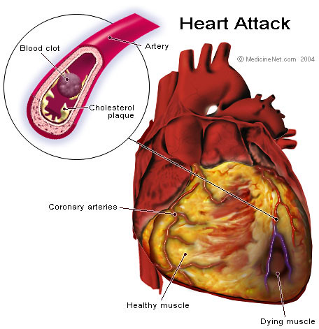
Gandhi, Ravindra. Heart Attack Anatomy. Digital image. Flickr. N.p., 11 May 2007. Web. 8 Nov. 2016.
Atherosclerosis, which is the depositing of plaque on the walls of blood vessels, is a precursor to both strokes and heart attacks and strokes, and is often caused by the ingestion of too much LDL and not enough HDL. This can cause problems when the plaque inside blood vessels blocks blood flow, or breaks off and gets stuck faster down the vessel where it is thinner. To promote my own cardiovascular health, I could exercise more each day, as well as eating foods richer in HDL and cutting down on my LDL intake. Additionally, I could check if there is a history of heart disease within my family, to be more aware of the risks that I could have. I wish to learn more about why men and older people are more likely to have high blood pressure, and whether that is due to anatomical differences or just the general lifestyles of that demographic.

Strengths/Weaknesses


During this unit, some of my strengths were being more responsible when doing homework and being able to say that I did my best work when it was being checked off. Again, however, I was not as diligent with my studying and was unable to properly space out my studying, so this will definitely be a point of focus for me for the rest of the semester. I think in terms of working with others, I definitely was able to gain a lot more insight into certain ideas, as well as absorbing the enthusiasm and knowledge of some of my group mates. One thing that I am still unsure about is the specific functions of the different white blood cells, as well as the specific functions and workings of the parasympathetic and sympathetic systems.

Monday, November 7, 2016

Heart Dissection Lab

1. The purpose of the pericardium is to protect the heart and surround it in a tight wrapping to prevent it from possibly tearing apart from the force of its beating. It also lubricates the outside of the heart to prevent friction.
2. Arteries seem to be substantially thicker and have ridges on the inside, whereas veins are smooth on the inside and much thinner.
3. The auricles serves as extensions of the atria to allow for more blood to fill the heart. Being that they feel very elastic when stretched, it probably expands to let much more blood in than would regularly enter the atrium.
4. The exterior appearance of the ventricles, at first sight, seem to be a darker tissue as compared to the atria. The atria also are packed with much more visceral fat than the ventricles.
5. The exit of the coronary sinus can be seen on the posterior side of the heart, and unfortunately was blocked to the eye by the massive amount of fat covering the area of the right atria.
The posterior view of the heart
The exit from the inferior vena cava was also cut off from our heart, but would have been located at the end of the extension of the superior vena cava.
The right atrioventricular valve, or the tricuspid valve is white in color, and has branches connecting it to the papillary muscles, which are presumably the chordae tendinae.
6. The tricuspid valve looks as such:
7. The anchoring of the heart valves by the chordae tendinae and the papillary muscle are important to heart function because it prevents the valve from caving in on itself, and allowing backflow of the blood to happen.
8. The white chordae tendinae can be seen, and there are ridges inside of the auricle. The endocardium of the left atrium is shiny and pale as compare to the myocardium that we cut into.
Cut inside the pulmonary vein
9. The function of the semi-lunar valves are to prevent artery blood from re-entering the heart.
10a. This might happen because that is where the inferior vena cava is, and backflow in that area might send unnecessary blood back to where it was coming from, which is the lower half of the body.
b. You might expect to see complications such as shortness of breath if valve disease occurs in the left side of the body, because the pulmonary veins and the pulmonary artery are located in that general area; lack of efficiency in these parts of the heart means that pathway of deoxygenated blood to the lungs as well as the oxygenated blood from the lungs is impaired as well, causing both the heart and the lungs to work harder.
11.The chordae tendinae look like cartilage, and the papillary muscle as expected creates a thicker amount of tissue to cut through. 
Cut into the aorta
12. The left side of the heart is much more spacious and also meatier in the myocardium surrounding the atria as compared to the right side of the heart, most likely because there needs to be more muscle to pump blood throughout the body as compared to just the heart.
13. On the right side of the heart, we can see the right ventricle, and on the left side, the left left ventricle. The interventricular septum and atrioventricular septum separate the two chambers as the meaty barrier in between. The epicardium layer is the outermost layer, with the myocardium being the reddish flesh in the middle, and the endocardium lining the inside of the 2 ventricles that can be seen in the photo. Other features are labeled on the photo.

Heart Video:
Extra Credit: